SOME TERMINILOGIES
- Immunogen: Substance capable of an immune response (humoral or cellular).
- Antigenicity: The property that allows a substance to combine specifically with antibodies or TCRs, whether or not they are immunogenic.
- Immunogenicity: The ability to induce humoral and/or cell-mediated immune response.
- Epitope: The small area of chemical grouping on the antigen molecule that determines specific immune response and reacts specifically with antibody is called an antigenic determinant or epitope.
- Paratope: The combining area on the antibody molecule, corresponding to the epitope (determinant), is termed paratope.
ANTIGENS
- Are usually protein in nature and sometimes polysaccharide.
- When introduced into a living animal evokes specific immune response, either by producing specific antibody or specially sensitized T cells or both.
- The antigen reacts specifically with the corresponding antibody in some detectable (observable) manner.
- Antigens has two properties:
- Immunogenicity-capacity to induce antibody formation.
- Immunological reactivity- capacity to react with an antibody.
TYPES OF ANTIGENS:
- Complete antigen or Immunogen
- Complete antigen or immunogen are able to generate immune response by themselves.
- They are high molecular weight proteins (10,000) but some are polysaccharides.
- Hapten or incomplete antigen
- Low molecular weight (less than 10,000), defective antigen,
- usually non-protein substance.
- Doesn’t have immunogenicity but retain immunological reactivity i.e. antigenic.
- Hapten becomes immunogenic by combining with carrier molecule.
- Heterophile antigen
- The same or closely related antigens may sometimes occur in different biological species, classes and kingdoms.
- These are known as heterogenetic or heterophile antigens.
- Antigens of different species cross react with each other. For example;
- Paul Bunnel Test-EBV with sheep RBC.
- Weil Felix reaction- Proteus OX2, OX19, OX K with rickettssia alkali stable polysaccharide.
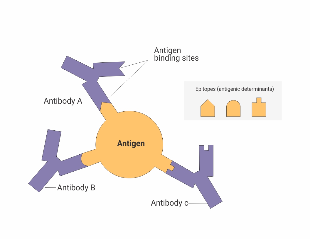
Antigenic determinants:
- Size: Larger size, more antigenic.
- Chemical: Decreasing order of antigencity-Protein> carbohydrate> lipid & nucleic acid.
- Susceptibility to tissue enzyme- increases antigenicity- (latex and d amino acid being chemically inert, are non antigenic)
- Foreignness- More the foreignness of the antigen, more is the antigenicity.
- Route of entry
- Genetic composition
- Specificity: Tissue specificity
Iso specific- Human RBC
Auto specific- HLA, Self antigens
Organ specific
ANTIBODIES
- Specialized serum proteins that are formed in response to an antigen and react specifically with that antigen or one very closely related to it some observable manner.
- Antibodies are synthesized by host B lymphocytes & plasma cells when they come in contact with a foreign antigen (e.g, infectious microbe).
- Chemically antibodies are globulins, hence are termed immunoglobulins.
FUNCTIONS
- Elimination (removal) of foreign antigen from the circulation.
- Recruitment and enhancement of host effector mechanisms.
- Measurement of specific antipathogen antibody levels in diagnosis.
- Passive administration of pooled antibodies for host therapy/protection.
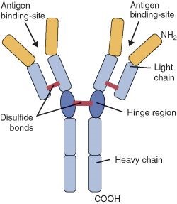
STRUCTURE OF IMMUNOGLOBULINS
- Immunoglobulin is Y shaped, consisting of identical 2 Heavy and 2 Light chains held together by disulfide (S-S) bonds.
- The H chains re structurally & antigenically distinct & contain constant and variable regions.
- Based on the structure of constant region of heavy chains, gamma (ϒ), mu (µ), alpha (α), delta (δ)& epsilon (ϵ), immunoglobulins are classified into IgG, IgM, IgA, IgD & IgE respectively.
- The L chains exist in 2 forms kappa (k) and lambda (λ).
- Each immunoglobulin molecule shows either 2 kappa or 2 lamda light chains but they are never found in combination.
- Papain digestion of immunoglobulin produces-
- 2 Fab portion-antigen binding site
- 1 Fc portion-perform biological function like complement attachment site, adherence to monocyte.
Properties of immunoglobulins
IgG
- Major immunoglobulin (70-80% of total), monomer consists of 2L & 2H chains.
- It has 4 subclasses; IgG1, IgG2, IgG3 & IgG4.
- Appears late usually after 2 weeks of infection and persists longer ( involves in secondary response).
- Takes part in precipitation, complement fixation & neutralization of toxin, viruses.
- Only immunoglobulin that can cross placenta.
- It has the longest half life-23 days.
IgA
- Principal immunoglobulin that appears in sero-mucous secretions such as saliva, tears, nasal fluids, sweat, colostrum, & secretions of the lungs, genitourinary & gastrointestinal tract.
- It occurs in 2 forms: Serum IgA & Secretory IgA.
- Protects exposed mucus membrane from microorganisms (local immunity).
- IgA in secretion is dimer while serum IgA is monomer.
- IgA is largely synthesized by plasma cells, only little amount may be derived from serum.
- Half life-6-8 days.
IgM
- Pentamer
- It has 10 antigen binding site.
- Mainly found in serum.
- Appears early in response to acute infection (involves in primary immune response).
- Aoffers protection against bacterimia.
- It is most efficient Ab that takes part in agglutination, CFT, & cytolytic action.
- It is a short lived immunoglobulin that disappears rapidly, its demonstration in serum indicates recent infection.
- Half life- 5 days
IgD
- <1% of the total immunoglobulin.
- Monomer consist of 2H & 2L chains.
- Mainly intravascular distribution.
- IgD & IgM occur on the surface of unstimulated B lymphocyes & serve as a recognition receptors for antigens.
- Combination of cell-membrane bound IgD or IgM with corresponding Ag leads to specific stimulation of the B cell-either activation, cloning to produce Ab or suppression.
- Half life-3 days.
IgE
- Also known as cytophilic antibody.
- Heat labile
- It has affinity for surface tissue cells , particularly mast cells & basophils.
- It mediates type I hypersensitivity reaction
- Increased IgE level is seen in parasitic (helminthic) reaction.
- Half life-2-3 days
ANTIGEN ANTIBODY REACTION
- Antigen antibody reaction is specific
- Entire molecules, not fragments react.
- Ag or Ab don’t denature during reaction.
- Combination occurs on the surface.
- Combination is firm but reversible (firmness of the union is influenced by the affinity and avidity of the reaction)-Bound together by weaker bonds such as ionic bond, Vander Waal’s force and hydrogen bond rather than stronger bond like covalent bond.
Affinity: denotes intensity of attraction between the antigen and antibody molecules.
Avidity: is the binding strength of individual antibody with its specific antigenic
determinants as a result of multivalent binding of antibody to antigen.
- Ag and Ab can combine in varying proportion, but optimum reaction at equilibrium (Marrack’s hypothesis).
USES
- In the body, they form the basis of antibody mediated immunity in infectious diseases or of tissue injury in some types of hypersensitivity and autoimmune diseases.
- In the laboratory, they help in diagnosis of infections.
- In epidemiological surveys, they assist in the identification of infectious agents and non-infectious agents such as enzymes & in screening the population for a particular infection.
- In general, these reactions can be used for detection & quantitiation of either antigens or antibodies.
Note: Ag-Ab reactions in vitro are known as serological reactions.
STAGES
- Primary stage:
In primary or initial interaction there is no visible effect & the reaction is rapid. However, microscopic localisation of antibody can be observed in a component of particular microorganism when the antiserum is labelled with a fluorecent dye.
- Secondary stage:
The primary interaction in most instances is followed by demonstrable events (secondary reaction), such as precipitation, agglutination or alternatively in the activation of nonantibody component such as serum complement or histamine from mast cells.
- Tertiary stage:
Some Ag-Ab reactions occurring in vivo initiate chain reactions that lead to neutralisation or destruction of injurious antigens, or to tissue damage.
These are tertiary reactions & include humoral immunity against infectious diseases as well as clinical allergy and other immunological diseases.
Principle-Marrack’s (Lattice) Hypothesis
- Multivalent Ag combines with bivalent Ab in varying proprtion depending on theAg-Ab ratio in the reactive mixture.
- Zone of equivalence- Antigen antibody reaction takes place at its best when the numbers of antigen & antibody are equal to each other.
- Antigen antibody reaction doesn’t take place properly if antigen or antibody level is excess.
Prozone phenomena or antibody excess occurs in:
-Enteric fever (Salmonella Typhi)
-Brucellosis
-Leptospirosis
-Syphilis
Post zone phenomena (Antigen excess) occurs in-
-Cryptococcus

PRECIPITATION
Definition
- Soluble antigen+ antibody at a suitable temperature, pH and electrolytesàleads to formation of insoluble precipitate or floccules(Ag-Ab complex).
- When the precipitate remains suspended instead of sedimenting, the reaction is called flocculation.
- Precipitation reaction may occur in liquid medium or in semisolid medium like agar gels, agarose gels or polyacrylamide.
- Test may be carried out as qualitative or quantitative.
- It is sensitive in the detection of antigen and as little as 1 µg of protein can be detected.
- Less sensitive for detection of antibodies.
Has many applications:
- Forensic application in the identification of blood & seminal stains
- Testing for food adulteration
- Grouping of streptococci by the Lancefield technique
- The VDRL test for syphilis
- To standardise toxins and toxoids
- To test toxigenecity in diptheria bacilli
Simple precipitation test
Ring test
- Antigen solution is layered over a column of whole serum (antibody) in a narrow test tube.
- A ring of precipitate forms at the junction of two.
Uses:
- Ascoli’s thermoprecipitin test
- Streptococcal grouping (Lancefield grouping)
Slide test
- A drop of each antigen solution and serum is placed on the slide & mixed by shaking.
- Flocculates form.
Uses:
Tube test
- Serial dilution of antigen made in test tube to which fixed amount of serum is added.
- Amount of antigen that flocculates the serum is called Lf dose.
Gel diffusion test:
- Precipitation test done in agar gel is called gel diffusion or immunodiffusion.
- These offer sensitive and specific results.
- The reaction is visible in form of distinct band of precipitation & reactive bands can be stained for better viewing as well as preservation.
Types:
- Single diffusion in one dimension (Oudin Procedure)
- Double diffusion in one dimension (Oakley Fulthorpe Procedure)
- Single diffusion in two dimension (Radial immunodiffusion)
- Double diffusion in two dimension-E.g. Elek’s gel precipitation (C. diptheriae toxigenecity testing) and Eiken test (E. coli)
- Immunoelectrophoresis: It combines electrophoretic separation of a composite antigen (serum) into its constituents, followed by immunodiffusion against its antiserum, resulting in separate precipitin lines, indicating reaction between each individual protein with its antibody.
This enables identification and approximate quantification of the various proteins present in serum.
Countercurrent Immunoelectrophoresis:
- It relies on movement of antigen and antibody in opposite direction.
- Simultaneous, electrophoresis of Ag & Ab in gel is done in opposite direction resulting in precipitation at a point between them.
- Used for detection of Alpha Feto Protein, Antigen of Cryptococcus and Meningococcus.
Rocket electrophoresis: one dimensional single immunoelectrophoresis
- The main application is for quantitative estimation of antigens.
Laurell’s two dimensional immunoelectrophoresis
- By this method, one can quantitate each of several antigens in a mixture.
AGGLUTINATION
Definition
- Insoluble antigen+ antibody at a suitable temperature, pH and electrolytesàleads to clump formation.
Types of test Procedure:
Slide/tile agglutination test:
- Provisional or qualitative procedure.
- The cells (living/dead bacteria, red cells etc.) are suspended in a drop of saline on a slide & a small drop of antiserum is added.
- This is rotated for a minute & presence or absence of clumping is noted.
Examples:
- ASO for Streptococcal infection
- Coagglutination test for staphylococcus.
- CRP
Tube Test:
- More quantitative than slide agglutination test.
Examples:
- Widal test for enteric fever
- Weil-Felix reaction for Typhus fever.
- Paul Bunnel test for infectious mononucleosis (Direct active haemagglutination)
- Coombs test (Indirect active haemagglutination test)
- TPHA test for syphilis (Direct passive haemagglutination test)
- Rose waller test for rheumatoid arthritis (Indirect passive haemagglutination test).
Application of Agglutination
- Identification of unknown culture.
- Blood grouping and cross matching.
- Serological diagnosis of enteric fever (Widal test), typhus (Weil-Felix reaction), Brucellosis and infectious mononucleosis (Paul Bunnel test).
- Streptococcus MG agglutination test for diagnosis of primary atypicla pneumonia
COMPLEMENT FIXATION TEST
- The abilty of Ag-Ab complexes to fix the complement is used in CFT.
- It is a versatile & sensitive test applicable with a variety of Ags & Abs & capable of detecting as little as 0.04 mg of Ab nitrogen & 0.1 mg of Ag.
- CFT is a complex procedure consisting of 2 steps and five reagents-antigen, antibody, complement, sheep erythrocytes & amboceptor (rabbit antibody to sheep red cells).
- Each of these reagents has to be separately standardised.
Uses: Wasserman test (syphilis), CFT for viral disease & other infectious diseases.

NEUTRALIZATION
Homologous Abs are able to neutralize the biological effects of viruses, toxins & enzymes. Such Abs are known as neutralizing Abs & the test is known as neutralization test.
- Toxin antitioxin neutralization test- When antitoxin combines toxin, it neutralizes biological effects of toxin making it harmless.
- Schick test-Diptheria toxin
- Naegler reaction
- Streptolysin O neutralization test
- Viral neutralization test- Virus neutralization can be demonstrated in various systems such as animals, tissue culture & chick embryos.
- Bacteriophage neutralization test-By plaque inhibition test.
OPSONIZATION
- Enhanced phagocytosis by coating the microbial surfaces by opsonins.
- Examples of opsonin molecules include:
- Antibodies: IgG and IgM
- Components of the complement system:C3b, C4b and iC3b
- Mannose binding lectin (initiates the formation of C3b)
- Most important components for opsonization-C3b & Fc (IgG)
Newer methods using labelled molecules
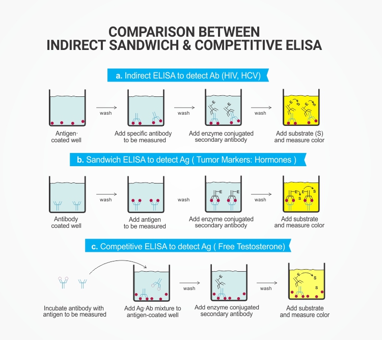
- ELISA (Enzyme Linked Immunosorbent Assay)-Enzyme tagged.
- RIA (Radioimmuno assay)-Radioactive isotope labelled.
- CLIA (Chemiluminoscent Linked Immunoassay)-Chemiluminoscent component like acridium
- IFA (Immunofluorescence assay)- Fluorescent dye labelled
ELISA
Enzyme-linked immunosorbant assay (El.lSA) is a non-isotopic immunoassay. An enzyme is used as a label in ELISA in place of radioactive isotope employed in RlA. ELISA is as sensitive as or even more sensitive than RlA.
ELISA is based on the immunochemical principles of antigen-antibody reaction.
- The antibody against the protein to be determined is fixed on an inert solid such as polystyrene.
- The biological sample containing the protein to be estimated is applied on the antibody coated surface.
- The protein antibody complex is then reacted with a second protein specific antibody to which an enzyme is covalently linked. Peroxidase, amylase and alkaline phosphatase are commonly used.
- After washing the unbound antibody linked enzyme, the enzyme bound to the second antibody complex is assayed.
- The enzyme activity is determined by its action on a substrate to form a product (usually coloured). This is related to the concentration of the protein being estimated.
Application of ELISA:
- HIV detection
- Infcetious diseases like hepatitis, EBV, cytomegalovirus IgM/IgG, dengue IgG, Influenza, TORCH panel etc.
- Rotavirus detection in fecal specimens and enterotoxin of E.coli in feces.
- Syphilis IgG/IgM, H. pylori IgG and antigen detection.
- Food toxins like chloramphenicol, streptomycin, peniciilin, aflatoxins, etc.
- Food adulteration include E.coli, campylobacter & Salmonella antigens.
- Mycobacterial antibody detection in tuberculosis.
- Human allergen-specific IgE and IgA ELISA.
Radioimmunoassay (RIA)
- Radioimmunoassay combines the principles of radioactivity of isotopes and immunological reactions of antigen and antibody, hence the name.
- It is an immunological assay to analyze any antigen or anti-body in the patient’s serum to diagnose the disease.
- It is one of the most sensitive & specific methods of immune assays available.
- It involves competitive binding of radio-labeled antigen and unlabeled antigen to a high-affinity antibody.
- The sensitivity range is 0.0006–0.006 µg antibody/ml.
It involves three principles which make it most specific & sensitive than other immune assays.
- An immune reaction i.e. antigen, antibody binding.
- A competitive binding or competitive displacement reaction. (It gives specificity)
- Measurement of radio emission. (It gives sensitivity)
Application of RIA
- RIA can estimate wide range of substances.These include peptides, steroid hormones, vitamins, drugs, antibiotics, nucleic acids, structural proteins and hormone receptor proteins.
- Radioimmunoassay has tremendous application in the diagnosis of hormonal disorders, cancers and therapeutic monitoring of drugs, besides being useful in biomedical research.
CLIA
- CLIA refers to a chemical reaction emitting energy in the form of light.
- Chemiluminescent compounds such as luminol or acridinium esters are used in CLIA as the label to provide the signal during the Ag-Ab reaction.
- The signal (light) can be amplified, measured & the concentration of the analyte calculated.
- The method is fully automated and handy in laboratories where work volume is large.
ICT (Immunochromatographic Test)
- Also known as Rapid test/dipstick or strip test or lateral flow assay.
- Uses Nitrocellulose membrane (NCM).
- Wide application due to its simplicity, economy, & reliablity.
- Test is claimed to be as sensitive & specific as EIA tests.
- Examples: Malaria Ag detection test, HIV Ab detection kits, HBsAg detection kits.
Newer Methods Using Nitrocellulose Membrane
Immunoblotting:Combines the sensitivity of the enzyme immunoassay with much greater specificity.
3 stages-
- PAGE (Polyacrylamide gel electrophoresis)àElectrophoretically mobilize and separate the antigen fragments
- Nitrocellulose membrane blottingà antigenic fragments are blotted on NCM
- Enzyme immunoassay-to detect the antibodies in serum of the patient.
- Nitrocellulose sheets with transferred proteins are called Western blot.
---The western blot test is the confirmative test for serodiagnosis of HIV infection.
- Nitrocellulose sheets with transferred DNA are called Southern blot.
- Nitrocellulose sheets with transferred RNA are called Northern blot.